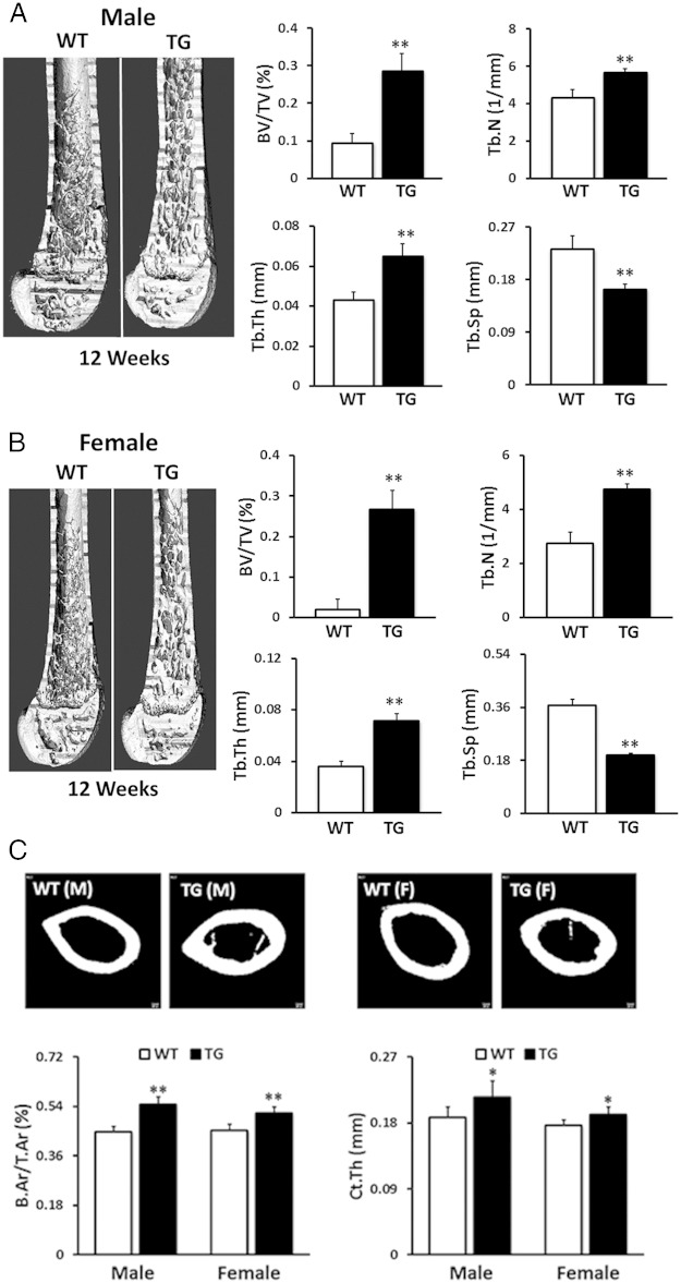
Figure 3.
Trabecular and cortical bone morphometry measured by μCT. Compared with WT mice, male (A) and female (B) TG WNT16 mice showed significantly higher trabecular bone volume (BV/TV), Tb.N, and Tb.Th but significantly lower Tb.Sp at distal femur at 12 weeks of age. Cortical bone morphometry at the femur midshaft revealed that both male and female TG WNT16 mice had higher cortical B.Ar/T.Ar and Ct.Th, compared with their WT littermates at 12 weeks of age (C); *, P < .005; **, P < .0001.

