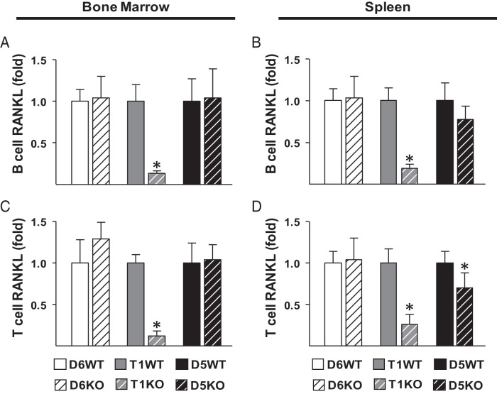
Figure 3.
RL-T1 and RL-D5 enhancer regions regulate RANKL expression in lymphocytes. A–D, B cells (A and B) and T cells (C and D) were isolated by positive selection from bone marrow (A and C) and spleens (B and D) of 4- to 6-month-old male D5KO, D6KO, and T1KO mice and their littermate controls. RANKL mRNA levels of the isolated lymphocytes were measured by RT-PCR analysis. The values represent the means ± SD of 3 to 8 mice/group. *, P < .05 vs littermate WT group by t test.

