Figure 6.
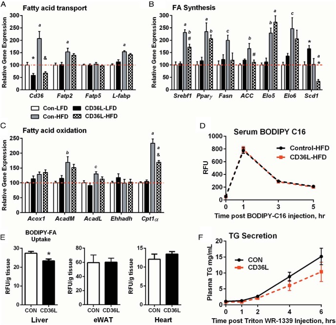
Hepatic BODIPY-FA-C16 uptake is impaired in CD36L mice on a HFD. A–C, Hepatic gene expression patterns for FA transport (A), FA synthesis (B), and β-oxidation genes (C) normalized to CON-LFD expression (n = 5–9/group). *, P < .05, #, P < .01, &, P < .001 compared with CON on same diet; c, P < .05, b, P < .01, a, P < .001 compared with same genotype. D and E, In vivo BODIPY-FA-C16 uptake was performed in CON and CD36L mice on HFD for 6 weeks. D, Serum BODIPY-FA-C16 fluorescence at indicated time points. E, Liver, epididymal WAT (eWAT) and cardiac tissue BODIPY accumulation normalized to tissue weight (n = 6–7 per group). **, P < .01 compared with CON. F, TG secretion assay was performed in CON and CD36L mice on a 22% fat diet after a 4-hour fast (n = 10 per group). All data are shown as mean ± SEM.

