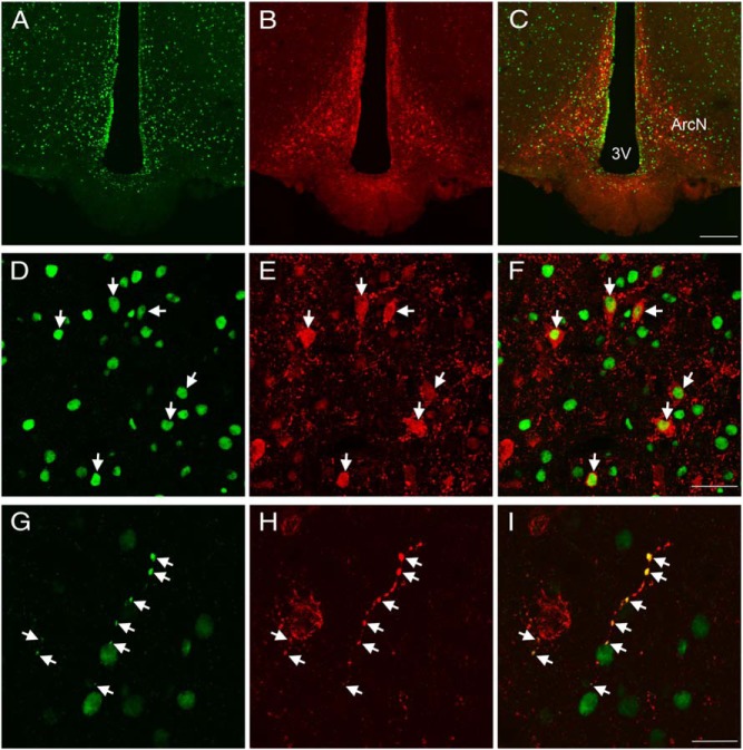
Figure 5.
Colocalization of EP24.15 and Kiss1 immunoreactivities in the ArcN of a metestrous female rat. Representative confocal photomicrograph of EP24.15 (green) (A) and Kiss1 (red) immunoreactivity in the ArcN (B). C, Merged image of A and B showing overlap and codistribution of Kiss1 and EP24.15. Numerous Kiss1 and EP24.15-immunoreactive cells are distributed throughout the ArcN (scale bar, 200 μm). Higher magnification of the ArcN demonstrating EP24.15 (D) and Kiss1 immunoreactivity (E). F, Colocalization of EP24.15 and Kiss1 in the ArcN. Arrows highlight regions of colocalization (scale bar, 40 μm). G, Colocalization of EP24.15 and H. Kiss1 in a nerve fiber. I, Merged image of G and H demonstrating coexpression along a “beaded” fiber (scale bar, 20 μm). 3V, third ventricle.

