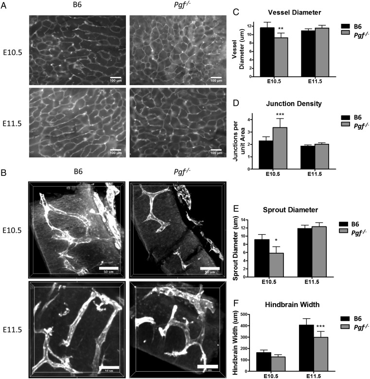
Figure 4.
Whole mount vascular staining of hindbrain (HB) at embryonic day (E) 10.5 and 11.5. HB vascularization was compared between B6 and Pgf −/− fetuses at E10.5 and 11.5 by examining the isolectin B4 (IB4) stained (white) ventricular plexus (A) and sprouting in transverse sections of the hindbrain (B). In the ventricular plexus, vessel diameter was thinner in the Pgf −/− HB vasculature at E10.5 (C). Conversely, the number of junctions per area was significantly greater in the E10.5 Pgf −/− HB vascular plexus (D). Confocal imaging of the HB cross-sections revealed smaller sprout diameter (E) and reduced HB thickness (F) in the Pgf −/− HBs. Means with 95% confidence intervals are shown with P < 0.05, P < 0.01, and P < 0.001 represented by *, **, and *** respectively.

