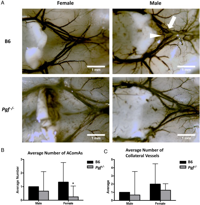
Figure 6.
Ink perfusion and imaging of the adult circle of Willis. The circle of Willis was imaged in adult male and female B6 and Pgf −/− mice (A). The average number of anterior communicating arteries (AComAs) (arrowhead) was decreased in female Pgf −/− mice (B). There was no significant difference in the number of anterior collateral vessels on the anterior cerebral arteries (arrow) in either male or female Pgf −/− mice (C). Means with 95% confidence intervals are shown with P < 0.05 represented by *.

