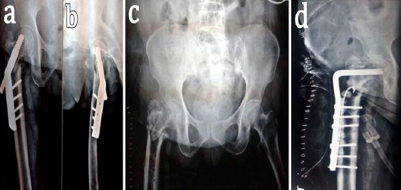
Figure 6.

Post-DHS infection in a 67-year-old female. (a and b) Antero-posterior and lateral view radiographs showing backing out of the Richard screw with non-union at the fracture site. (c) Radiograph of pelvis post implant exit showing non-union at the fracture site. (d) Antero-posterior radiograph showing condylar blade plate fixation of the infected non-union with bone grafts and antibiotic beads in-situ.
