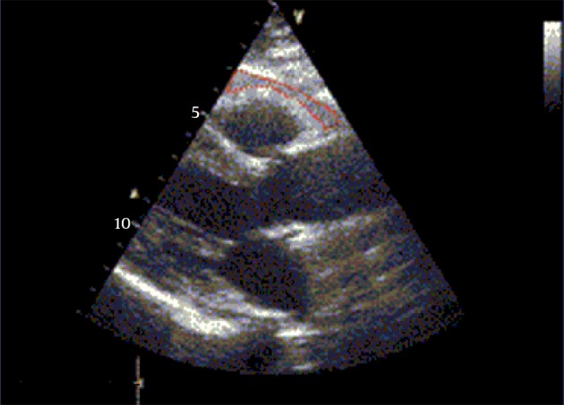
Figure 1. Echcocardiographic Epicardial Adipose Tissue Thickness.

Whit in red dashed shape is identified as the echo free space between the outer wall of the myocardium and the visceral layer of the pericardium in the parasternal long axis view.

Whit in red dashed shape is identified as the echo free space between the outer wall of the myocardium and the visceral layer of the pericardium in the parasternal long axis view.