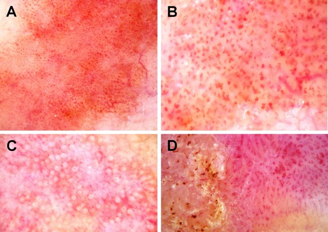
Figure 1.
Videodermoscopy of squamous cell carcinoma in situ. 70-fold magnification. (A) SCC in situ with small dotted vessels distributed in packed clusters; (B) SCC in situ with glomerular vessels; (C) SCC in situ with white circles; (D) SCC in situ with dotted and glomerular vessels, and hyperkeratosis.

