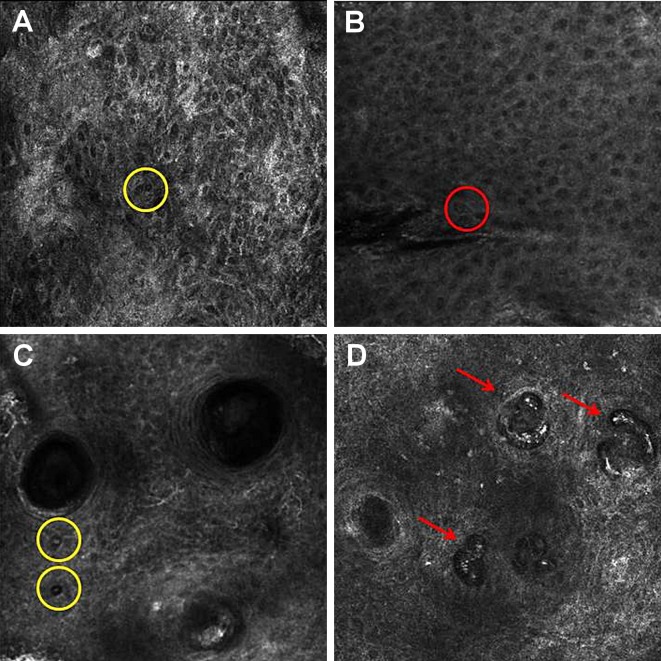
Figure 2.
Reflectance confocal microscopy of squamous cell carcinoma. (A) SCC with a disarranged pattern of the spinous-granular layer of the epidermis and round cell with a bright center and a dark peripheral halo (yellow circle); (B) SCC with an atypical honeycomb and round cell with a dark center and a bright rim surrounded by a dark halo (red circle); (C) SCC with an atypical honeycomb and round cell with a bright center and a dark peripheral halo (yellow circle); (D) SCC with round blood vessels in the dermis.

