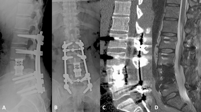
Fig. 2.

Postoperative sagittal (A) and anteroposterior (B) X-ray films, and sagittal noncontrast computed tomography (C) images demonstrating L4 vertebral body resection with expandable cage reconstruction and pedicle instrumentation from L2 to S1 with cross-link placement at L2–3 and L5–S1. (D) Sagittal T2-weighted noncontrast magnetic resonance image demonstrating resection of the L4 vertebral body with decompressive laminectomy and evacuation of epidural abscess.
