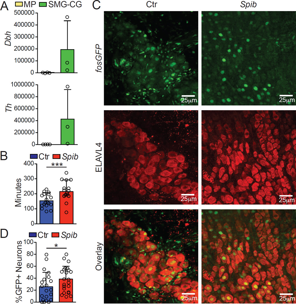
Figure 6. Extrinsic Sympathetic Innervation is Activated During Enteric Infection.
(A) Expression of polysome-associated mRNA from small intestine muscularis and SMG-CG of naïve Snap25RiboTag mice determined by qPCR, presented relative to Hprt. (B) Total GI transit time (time required to expel feces containing carmine red dye) in mice 30 min post intragastric exposure to Spib or PBS (control). (C) Confocal images from the SMG-CG isolated from FosGFP mice 2 h post Spib intragastric exposure and stained using anti-GFP (upper panels) and anti- ELAVL4 (middle panels) antibodies. (D) Quantification of the number of GFP+ nuclei amongst ELAVL4+-TH+ neurons within SMG-CG. (A), pooled data shown are from 3 independent experiments, n = 3; (B), pooled data shown are from 4 independent experiments, n = 18; (C), images are representative of 2 independent experiments, n=6; (D), pooled data shown are from 2 independent experiments, n = 6 (each dot represents one image of sympathetic ganglion neurons). Data were analyzed by unpaired T-test and are shown as average±SD, *p ≤ 0.05,***p ≤ 0.001. See also Figure S4.

