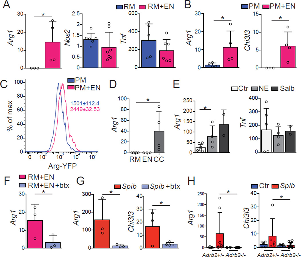
Figure 7. β2ARs–Mediate Polarization of Macrophages.
(A–F) WT mice neuro-sphere–derived primary enteric-associated neurons (EANs) were cocultured with RAW (RM) or peritoneal macrophages (PM) from WT mice. (A, B) Expression of mRNA for Arg1, Nos2, Tnf and Chi3l3 by sorted (A) RM or (B) PM 18 h post co-culture with EANs. (C) Representative flow cytometry histogram for YFP expression by sorted PM isolated from Arg1YFP mice cultured with EANs as in A. (D) Expression of mRNA for Arg1 by sorted RM 18 h post exposure to conditioned media from RM, EANs or RM-EANs co-cultures (CC). (E) Expression of mRNA for Arg1 and Tnf by sorted PM 1 h post exposure to NE or Salbutamol (β2AR agonists). (F) Expression of mRNA for Arg1 by sorted RM 18 h post co-culture with EANs with or without butaxamine (β2AR-selective blocker). (G) Expression of mRNA for Arg1 and Chi3l3 by sorted small intestine MMs isolated 2 h post intragastric exposure to Spib in mice treated with vehicle or butaxamine. (H) Expression of mRNA for Arg1 and Chi3l3 by sorted small intestine MMs isolated from Adrb1+/−Adrb2−/− and Adrb1+/−Adrb2+/− littermate control mice 2 h post intragastric exposure to Spib. qPCR results are presented relative to Rpl32 expression. (A-H), pooled data of at least 2 independent experiments (A, n = 3–7; B-D, n = 3–4; E, n = 2–4; F and G, n = 3–4;H, n = 4–11. Data were analyzed by unpaired T-test and are shown as average±SD, *p ≤ 0.05. See also Figure S2.

