
Figure 6.
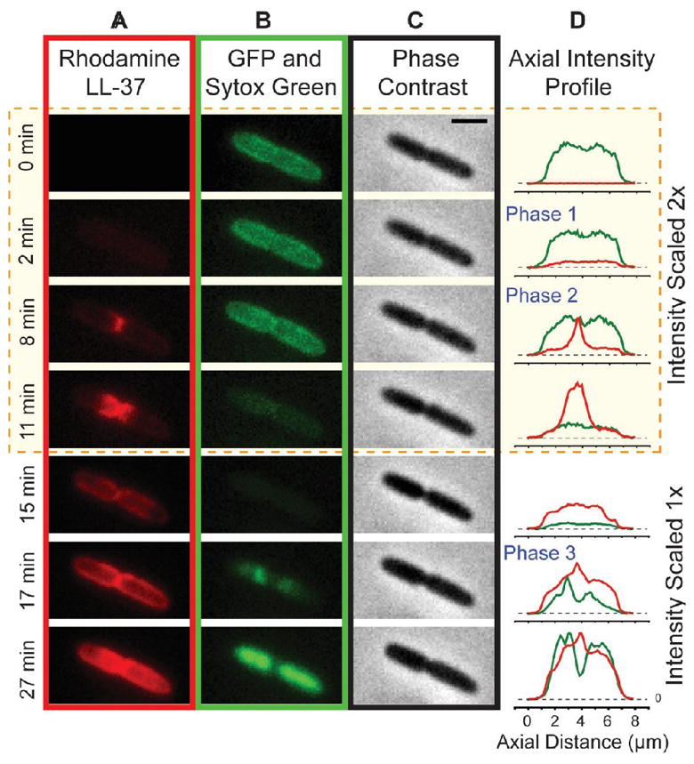
Attack of Rh-LL-37 on a Septating E. coli Cell. Montage at left shows time sequences of red (A; Rh-LL-37), green (B; first periplasmic GFP, then Sytox Green), and phase contrast images (C). (D) Axial projections of red and green intensity distributions. Reproduced from [29] with permission.