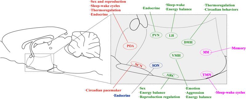
Fig. 1.
An overview of hypothalamic nuclei and functions. Nuclei and their functions in the preoptic region are denoted in red, including the preoptic area (POA) and the suprachiasmatic nucleus (SCN). Relative locations and functions of the supraoptic region are denoted in blue, including the supraoptic nucleus (SON). The tuberal region is denoted in green, including the arcuate nucleus (ARC), the paraventricular nucleus (PVN), the lateral hypothalamus (LH), the ventromedial hypothalamus (VMH), and the dorsomedial hypothalamus (DMH). The mammillary region, including the mammillary area (MM) and the tuberomammillary nucleus (TMN), is denoted in purple

