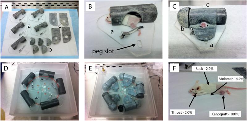
Fig. 1.
Schematic of the mouse irradiation device and shielding efficiency. A: Individual mouse treatment chambers and component parts: (a) primary lead-shielded chamber with low-pressure left leg clamp; (b) lead head caps; and (c) lead flank shields with pre-cut circular collimation. B: Initial anesthetized mouse positioning within the body chamber with leg clamp engaged. C: Fully shielded individual chamber, with circular cut-out collimation for left flank/leg xenograft exposure. D: Five-chamber arrangement (with mice and components not included). The red circle identifies the edge of the beam cone. The small red dots are the tops of the pegs for position reproducibility. Xenografts were aligned equidistant from the beam center and within the cone to ensure a uniform dose-rate (approximately 285 cGy/minute) and total dose delivery per fraction. E: Complete set-up prior to treatment. The semi-sealed treatment box with inhalational anesthesia gas tubing attached to lid valve. F: Shielding quality assurance results based on body location. Measurements of doses received by the shielded, non-targeted animal, relative to the unshielded xenograft doses revealed 95.8–98.0% shielding for all anatomic sites.

