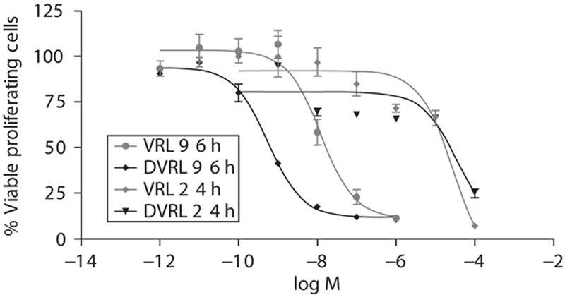
Fig. 2.

Inhibition of endothelial cell proliferation by VRL and DVRL. HUVECs were exposed to the indicated concentrations of VRL/DVRL for either 24 or 96 h. Viable cells were determined using an MTS assay. Results are expressed as a percentage of viable control cells and half-maximal inhibitory concentrations (IC50). DVRL, 4-O-deacetylvinorelbine; HUVEC, human umbilical vein endothelial cell; VRL, vinorelbine.
