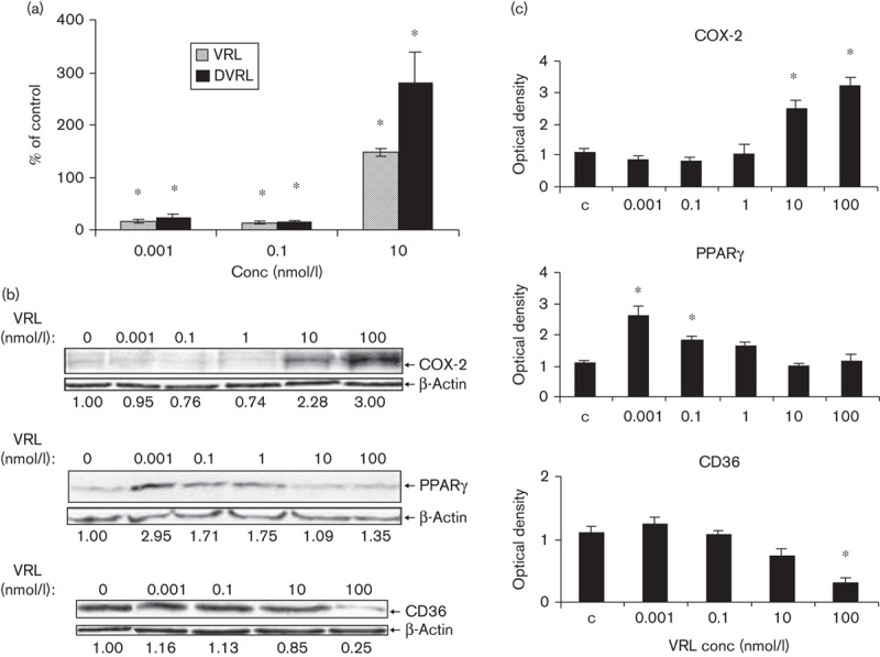
Fig. 5.

VRL/DVRL on angiogenic and antiangiogenic proteins in the protracted administration model. (a) IL-8 protein levels were determined by ELISA. (b) Protein levels of COX-2, PPARγ and CD36 were determined by western blotting using respective antibodies; the last blot shows only the nonglycosylated but full-length CD36 protein (55 kDa). The numbers under the blots indicate values normalized to β-actin values of the respective proteins. Representative figures of two independent experiments are shown. *P<0.05 compared with control group. COX, cyclooxygenase; DVRL, 4-O-deacetylvinorelbine; HUVEC, human umbilical vein endothelial cell; IL, interleukin; PPARγ, peroxisome proliferator-activated receptor γ; VRL, vinorelbine.
