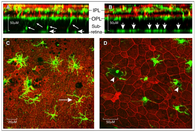
Figure 3. Retinal microglial in the aging eye.
A-B, Reconstructed z-stack confocal images from a 16-month (A) and 27-month (B) old mouse retina stained for Iba-1 (green for microglia) and lectin B4 (red for blood vessels). A, at 16 months, few Iba-1+ microglial cells were detected at the subretinal space (short arrows) and some were still connected to cells in the OPL layer (small arrows). B, at 27 months many more Iba-1+ cells were detected at the subretinal space (short arrows), and no Iba-1+ cells were detected between the OPL and subretinal space. C, heterogeneous morphology of Iba-1+ cells at the subretinal space in a 18-month old mouse. Most of the cells have larger cell bodies and shorter dendrites, and a few cells display a relatively small cell body and long dendrites (arrow). D, subretinal Iba-1+ cells from a 27-month old mice showing pigmented cell body (arrowheads). IPL – inner plexiform layer; OPL – outer plexiform layer.

