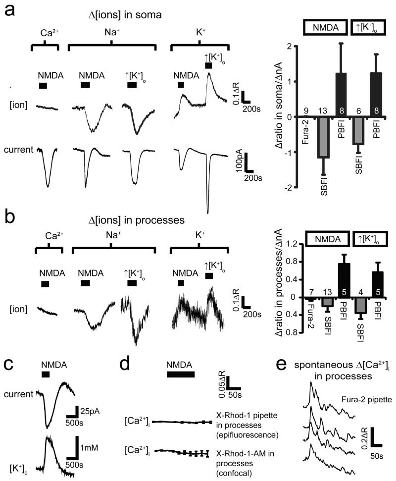
Figure 2. NMDA does not elevate [Ca2+]i in oligodendrocytes.
a-b Oligodendrocyte membrane current (lower traces, a) and background-subtracted fluorescent dye ratio (R, see Methods, concentration increases are upwards for all dyes) when measuring [Ca2+]i with Fura-2, [Na+]i with SBFI, and [K+]i with PBFI; 100μM NMDA was applied, or [K+]o was raised from 2.5 to 5 mM, with fluorescence measured in soma (a) or myelinating processes (b). Right panels: peak fluorescence change normalised to evoked current (number of cells on bars). c NMDA-evoked current and simultaneously recorded [K+]o. d Measuring [Ca2+]i with X-Rhod-1, loaded from pipette or as an acetoxymethyl ester2, reveals no NMDA-evoked [Ca2+]i rise. e Spontaneous [Ca2+]i transients in four myelinating processes confirm Fura-2 is working. Error bars, s.e.m.

