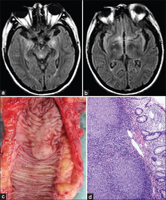
Figure 1.

Neuroimaging and pathological findings of the patient. Brain magnetic resonance imaging fluid-attenuated inversion recovery sequences showed abnormity in medial temporal lobe (a) and diencephale (b). Resected tissue of terminal ileum demonstrated a focal mass (c). Pathological findings of submucosal follicular lymphoma (d), H E staining, ×200.
