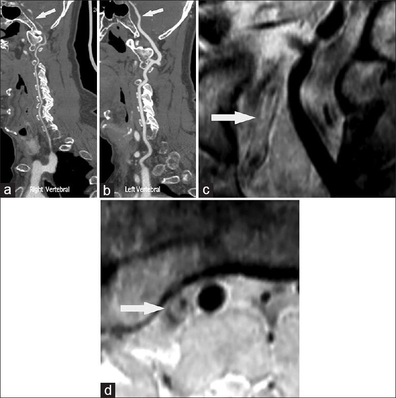
Figure 1.

Computer tomography angiography suggested right vertebral artery hypoplasia (a, arrow). The left vertebral artery (VA) was the dominant one (b arrow). Coronal and axil reconstructed views of high resolution magnetic resonance images (HR MRI) showed a large thrombus leading to lumen occlusion in the middle portion (c, arrow) and eccentric plaque in the distal portion of intracranial VA (d, arrow). This case was diagnosed as acquired atherosclerotic stenosis since HR MRI showed similar outer vessel diameter in the right VA as that in the left VA.
