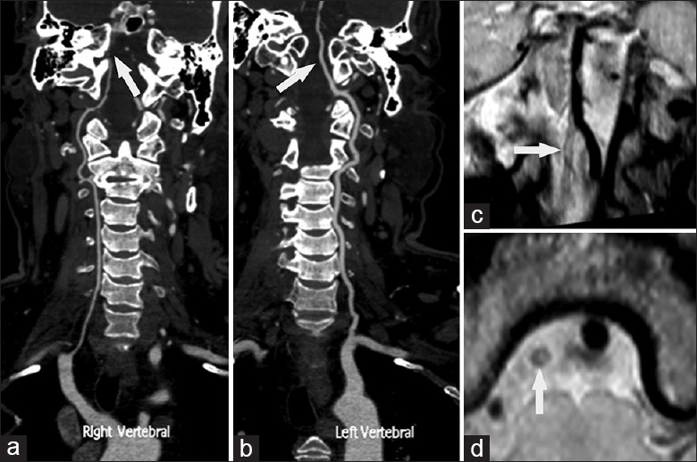
Figure 5.

Computer tomography angiography suggested right vertebral artery hypoplasia (VAH) with absence of lumen in the distal portion of intracranial vertebral artery (VA) (a arrow). The left VA was dominant side (b arrow). Coronal (c arrow) and axil (d, arrow) reconstructed views of high resolution magnetic resonance images showed wall thickening with a large thrombus leading to lumen occlusion in the distal portion of intracranial VA. High resolution magnetic resonance images showed outer vessel diameter in the right VA was less than 50% of the left VA. This case was diagnosed as VAH with atherosclerosis and thrombus.
