Figure 1.

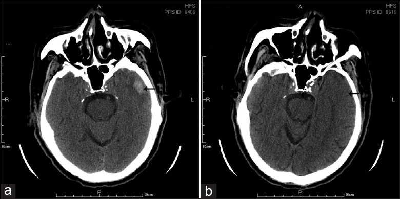
(a) Computed tomography image (CT) of a 20 mm × 11 mm intracranial hemorrhage in the left temporal lobe; (b) Follow-up CT brain obtained on 50th days revealed the disappearance of hematoma.

(a) Computed tomography image (CT) of a 20 mm × 11 mm intracranial hemorrhage in the left temporal lobe; (b) Follow-up CT brain obtained on 50th days revealed the disappearance of hematoma.