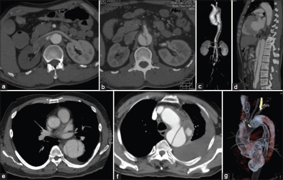
Figure 1.

Preoperative computed tomography angiography showed the “complicated” type B aortic dissection. Features of right renal malperfusion with great compression of true lumen (a), with superior mesenteric artery malperfusion (b), with lower limb ischemia (c), with multi-barrel and superior mesenteric artery malperfusion (d), with three barrels (e), with periaortic hematoma and hemorrhagic pleural effusion (f), and with a large tear (g) located in the proximal dissection near the left subclavian artery.
