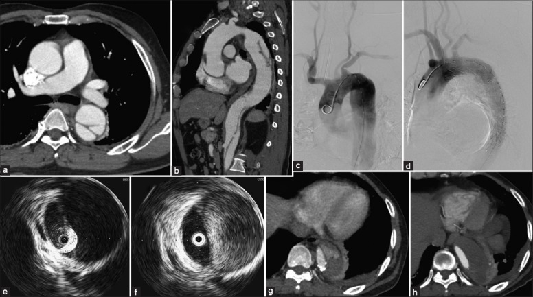
Figure 2.

(a and b) Preoperative computed tomography angiography in the intravascular ultrasound-assisted group (patient 11) demonstrated the diameter of the first entry tear was 9 mm and was 20 mm distal to the left subclavian artery. The false lumen located at the outer aortic curvature, whereas the true lumen with great compression at the descending aorta. Fortunately, the superior mesenteric artery generated from the true lumen. The left renal artery from the false lumen was not compromised; the right renal artery has thrombosed. (c-f) With the assistance of intraoperative intravascular ultrasound, the case was successfully repaired with a stent graft (c-d). The flap moved remarkably and the high velocity flow in the false lumen was observed in real-time by the intraoperative IVUS before (e) and after TEVAR (f). (g-h) Post-CTA at 13 months showed partial thrombosis at the end of the stent graft (g), whereas thrombosis was fully formed at 26 months follow-up (h).
