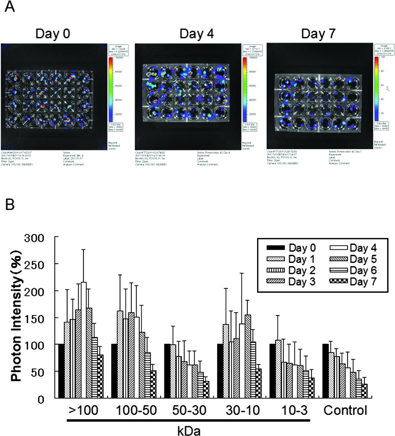
Figure 2.
Comparison of changes in luminescence intensity of islets in ET-Kyoto organ preservation solution after addition of medium conditioned with various fractions from mesenchymal stem cells (MSCs). (A) Photographs of Luc-Tg rat-derived islets in preservation solution treated with various fractions from the MSC-conditioned medium. From the left column on the plate, >100 kDa fraction, 50–100 kDa, 30–50 kDa, 10–30 kDa, 3–10 kDa, and 0 kDa (control). (B) Bioluminescence imaging using an in vivo imaging system to assess cell viability in the islets. Samples were exposed to extracellular-type trehalose-containing Kyoto (ET-Kyoto) organ preservation solution at 4°C. Data are representative of four independent experiments.

