Fig. 4.
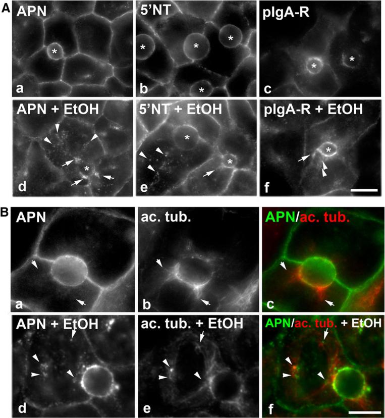
Transcytosing proteins are present on sub-canalicular structures that are aligned along acetylated microtubules in ethanol-treated cells. A, B Cells were treated in the absence (a–c) or presence of 50 mM ethanol (EtOH) (d–f) for 72 h as indicated. Live cells were labeled with the indicated canalicular proteins as described in Fig. 1, and the antibody-antigen complexes chased for 45 min. The transcytosing proteins are additionally detected in sub-canalicular clusters (marked with arrows) or discrete puncta (marked with arrow heads) in ethanol-treated cells (d–f). Bar 10 μm. In B, APN antibody-antigen complexes were continuously chased for 45 min and cells processed for immunofluorescence detection of both the trafficked APN and acetylated α-tubulin. Merged images are shown in c and f. Arrows are pointing to APN-positive structures that are immediately adjacent to acetylated microtubules in ethanol-treated cells. Bar 10 μm

