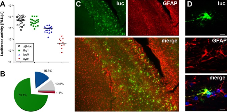
FIG 1.
Astrocytes are the main source of IFN-β during intracranial infection with TMEV. (A) Reporter mice in which the luciferase gene can be induced in all cell types (Δβ-luc), in astrocytes and neurons (thy1), in neurons only (syn1), or in microglia/macrophages (lysM) were infected with 104 PFU of TMEV GDVII, and luciferase activities in samples of brain homogenate were measured at day 3.5 postinfection. Background levels for uninfected mice were usually around 100 relative light units (RLU) per μl. (B) The average contributions to luciferase activity of different cell types are shown as pie charts. The mean activity of global Δβ-luc reporter mice was set to 100%. Green = astrocytes, blue = microglia/macrophages, red = neurons, gray = contribution of unidentified cells. (C) Luciferase-producing cells in the corpus callosum/hippocampus region were visualized by immunostaining. Most of the luciferase (luc)-producing cells were identified as astrocytes by costaining for GFAP. (D) A single luciferase-positive cell is shown at higher magnification to better visualize colocalization with GFAP. Bar = 10 μm.

