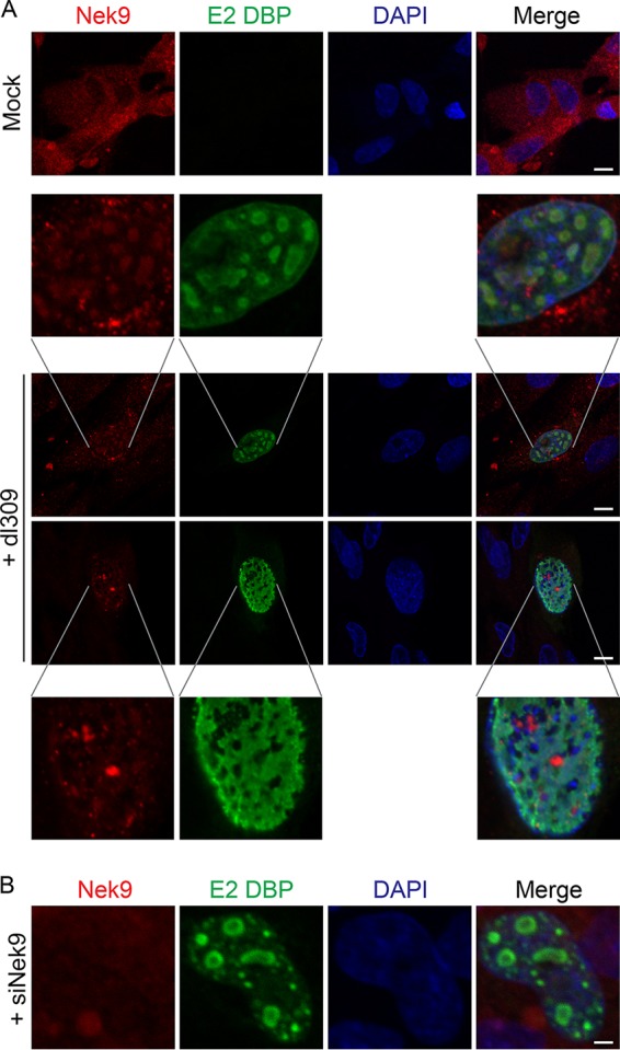
FIG 5.

Nek9 localizes to viral replication centers in infected cells. (A) IMR-90 cells were infected with dl309 at an MOI of 10, fixed, and stained for E2 DBP and Nek9 24 h after infection. Uninfected, control IMR-90 cells were also imaged, showing normal distribution of Nek9 (top panel). DAPI was used as a nuclear counterstain. Bar, 7.5 μm. (B) IMR-90 cells were initially transfected with siNek9 to deplete Nek9, infected with dl309 at an MOI of 5, and stained as described for panel A. Bar, 2 μm.
