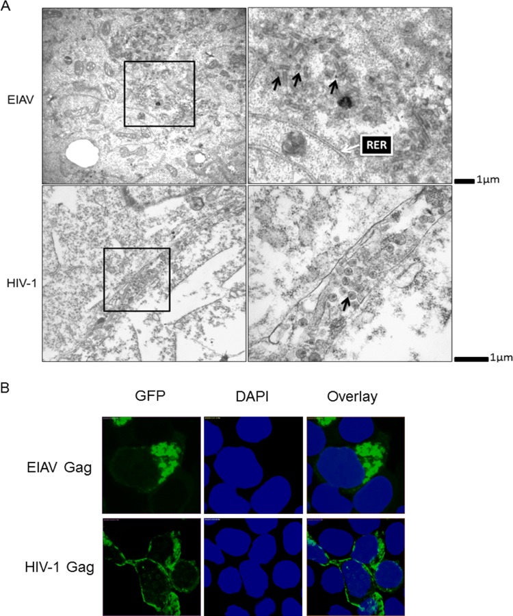
FIG 1.
Comparison of cellular localization of EIAV and HIV-1 viral particles. (A) EIAV VLPs were mainly localized at interior cellular membranes, while HIV-1 VLPs were detected at the plasma membrane. 293T cells were transfected with EIAV and HIV-1 Gag-Pol plasmids. After 48 h, the supernatants were discarded, and the cells were washed three times with PBS prior to harvesting for transmission electron microscopy. The intracellular VLPs are indicated by black arrows, and a typical RER that had a tubular appearance is indicated by a white arrow (28). (B) Cellular localization of EIAV and HIV-1 Gag were distinct. EIAV and HIV-1 Gag-GFP were transfected into 293T cells. After 48 h, the cells were fixed using formalin and stained with DAPI (blue). Gag-GFP expression and localization were analyzed by confocal microscopy.

