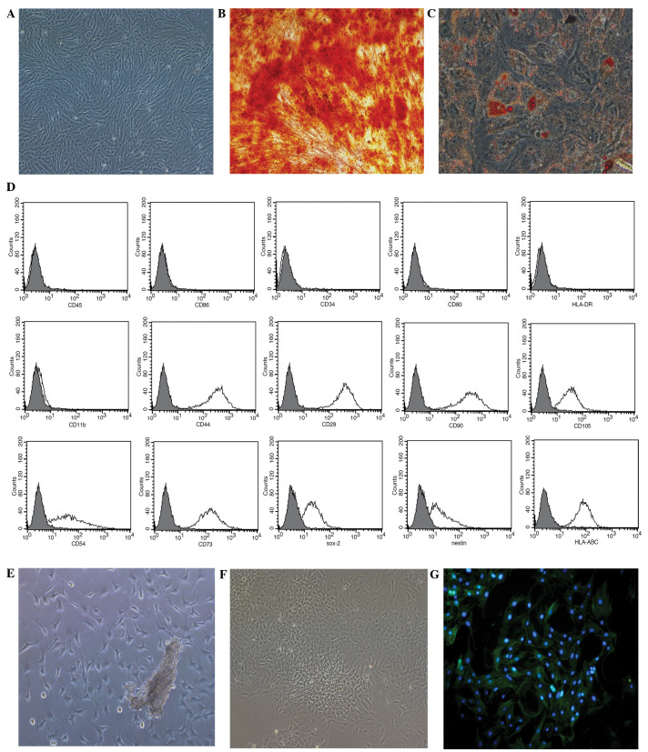
Figure 1.
Growth of mesenchymal stem cells (MSCs) and pulmonary artery smooth muscle cells (PASMCs) in vitro. (A) MSCs culured in serum-free medium at passage 3 (magnification, ×100). (B) Osteogenic and (C) adipogenic differentiation of MSCs cultured in MSC-conditioned media identified using Alizarin red S or Oil Red O stain, respectively (magnification, ×200). (D) Phenotypic analysis of MSCs, assessed using flow cytometry. (E) PASMC tissue explant cultured for 5 days (magnification, ×100). (F) PASMCs at passage 3 (magnification, ×100). (G) PASMC immunofluorescent staining for α-smooth muscle actin (magnification, ×200). CD, cluster of differentiation; Sox-2, SRY box-2; HLA, human leukocyte antigen.

