
Figure 3.
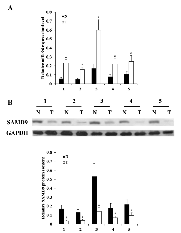
Expression of miR-96 and SAMD9 in NSCLC and adjacent normal lung tissues. The expression levels of (A) miR-96 and (B) SAMD9 protein in T and N lung tissues from 5 consecutive patients were determined with reverse transcription-quantitative polymerase chain reaction and western blot analysis, respectively. In western blot analysis, the density of the SAMD9 blots was normalized against that of GAPDH to obtain a relative blot density to indicate the relative SAMD9 protein content. *P<0.05 vs. N. NSCLC, non-small cell lung cancer; miR microRNA; SAMD9, sterile α motif domain-containing 9; T, NSCLC tumor; N, adjacent normal tissue.