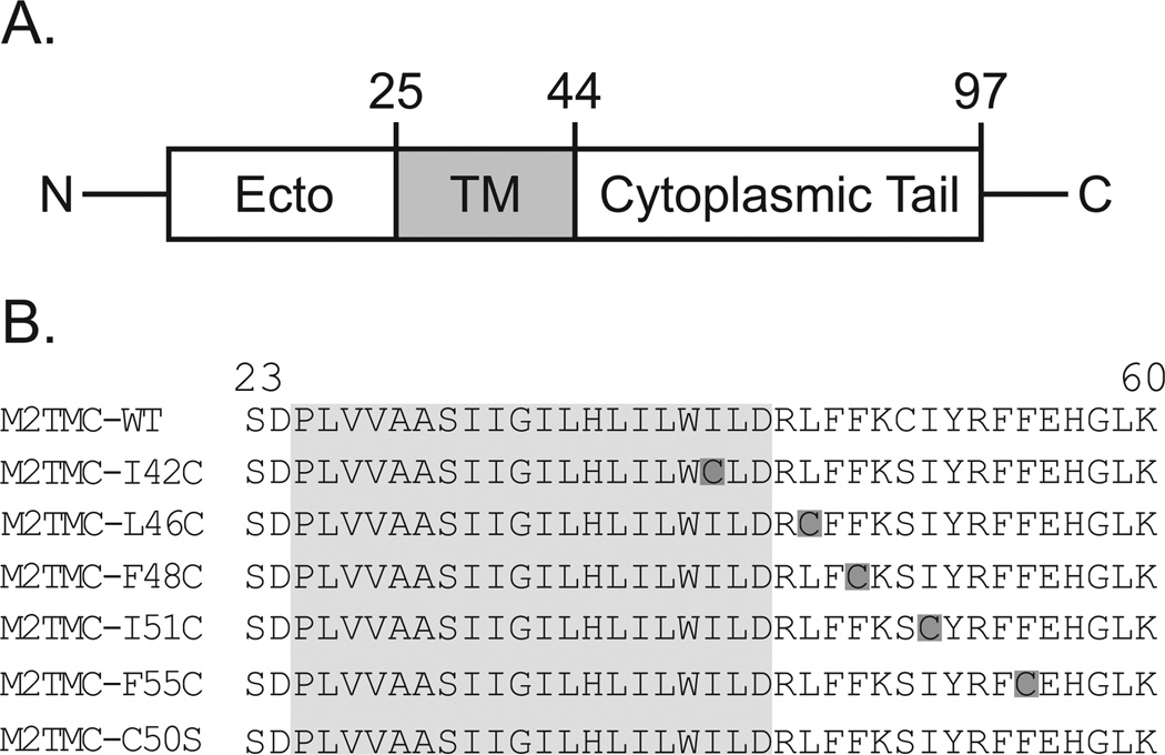
Figure 1. Spin-labeled M2 peptides used in this study.
A. Each monomer of the M2 protein is composed of a N-terminal ectodomain, a transmembrane domain (shaded in gray), and a C-terminal cytoplasmic domain. B. Wild-type sequence corresponds to the M2 protein from influenza strain A/Udorn/72 (H3N2). M2TMC (23–60) peptides were spin labeled at single cysteine sites (dark gray boxes). All M2TMC sequences other than the WT sequence have a C50S replacement in addition to the cysteine substitution necessary for spin labeling. The last cysteine-less sequence was used for spin dilution.

