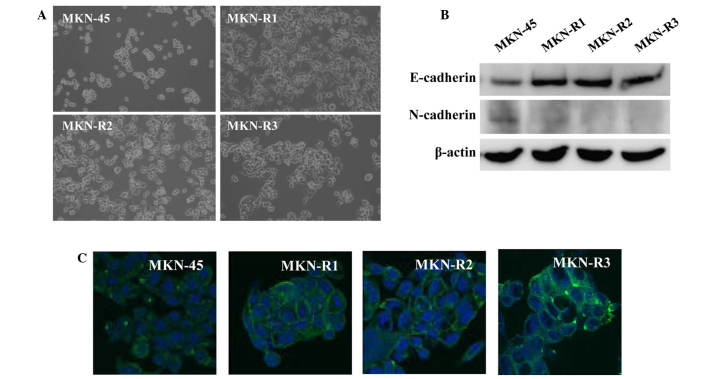
Figure 2.
Epithelial transition of MKN-45 cell line and clones resistant to KRC-108 (MKN-R1, -R2 and R3). (A) Phase contrast images of the MKN-45 and MKN-R cells (magnification, ×100). (B) The expression of E-cadherin and N-cadherin in the MKN-45 and MKN-R cells was detected by western blotting. (C) E-cadherin expression was analyzed by immunofluorescence: Green, E-cadherin; blue, DAPI (magnification, ×630).

