
FIG. 3.
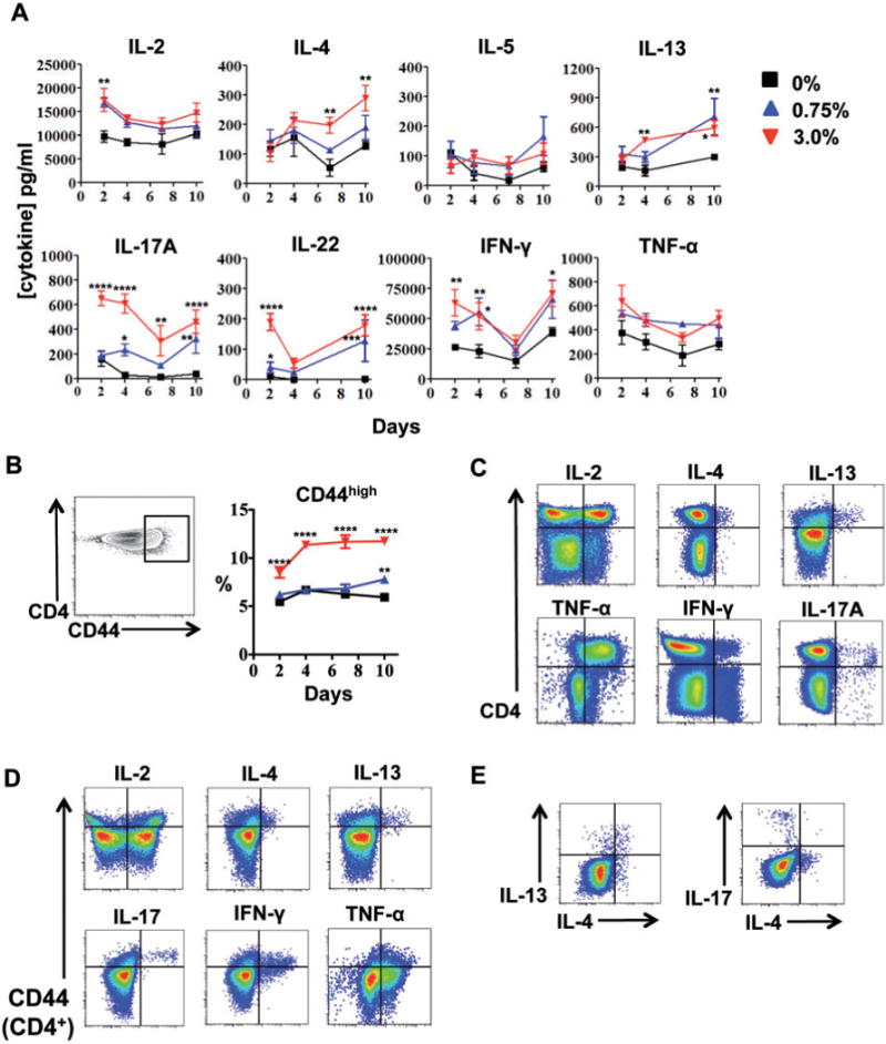
Repeated dermal exposure to triclosan augments cytokine production by activated LN CD4 T cells. Cytokine concentrations in LN cell supernatants (A) and percentage of LN CD4T cells expressing high levels of CD44 (B) from mice dosed daily with 0% (square), 0.75% (triangle) or 3.0% (inverted triangle) triclosan (n=4 mice) (A). Representative two-parameter histogram plots of LN CD4 T cell ICS staining from mice dosed daily with 3% triclosan and harvested Day 7 (C, D, E), versus CD44 expression (D) and in CD44+ CD4 T cells (E). Data are representative of 3 independent studies. Bars represent the mean ± SEM, with statistical significance indicated as P ≤ .05 (*), P≤ .01 (**), P≤ .001 (***), and P≤ .0001 (****).