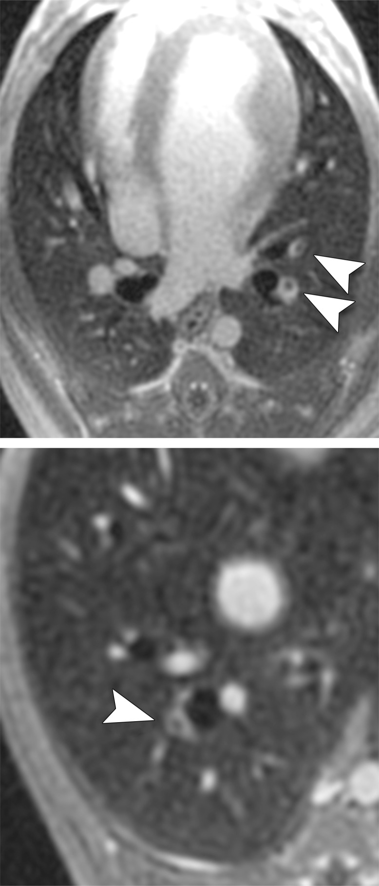
Figure 1b:

MR angiographic and UTE images used to detect PEs. (a) Breath-hold 3D MR angiographic, (b) free-breathing 3D radial UTE, and (c) CT images after induction of PE. Two segmental emboli (arrowheads in upper row) in the left caudal lobe and one subsegmental embolus (arrowheads in lower row) in the right caudal lobe were detected by both readers with both MR angiography and UTE imaging. UTE imaging shows high-resolution artifact-free depiction of the embolus and the lung parenchyma, including small bronchi within the same window setting.
