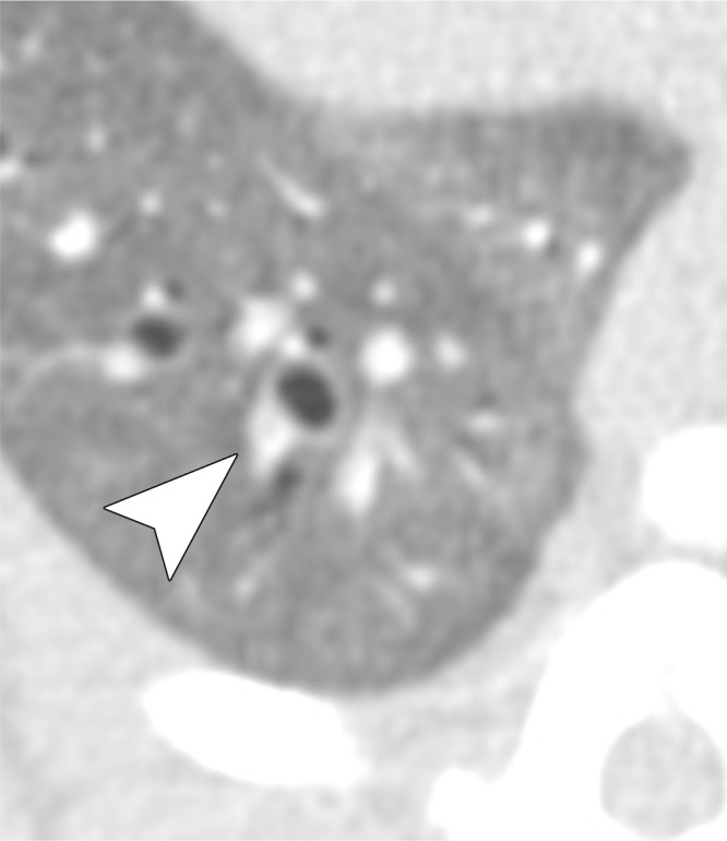
Figure 5d:

Examples of a subsegmental pulmonary embolus that was detected with UTE but not MR angiography. Axial (a) breath-hold MR angiographic, (b) free-breathing UTE, and (c, d) CT images displayed by using soft-tissue (c) and lung (d) window settings after induction of PE. Both readers detected a subsegmental embolus (arrowhead) in the right caudal lobe with UTE but did not detect the thrombus with MR angiography. Note the high-resolution and improved soft-tissue contrast of the lung parenchyma with UTE, allowing for depiction of the distal bronchus adjacent to the occluded artery.
