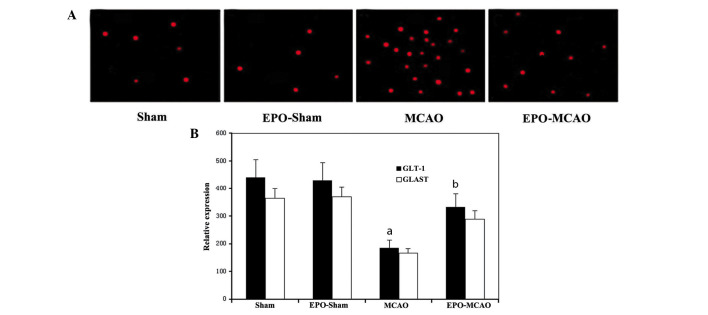
Figure 2.
Apoptotic neurons in rat brains were detected by TUNEL fluorescence double staining. (A) Representative TUNEL-positive apoptotic cells in the same infarct area of the ipsilateral cortex of the following groups: Sham (n=9), EPO-sham (n=8), MCAO (n=10) and EPO-MCAO (n=9). (B) Number of apoptotic cells per field in the same infarct area of each group. Data are presented as the mean ± standard deviation. aP<0.01 vs. the sham group; bP<0.05 vs. the MCAO group at the same reperfusion time. EPO, erythropoietin; MCAO, middle cerebral artery occlusion; GLT-1, glutamate transporter 1; GLAST, glutamate aspartate transporter; TUNEL, terminal deoxynucleotidyl transferase-mediated dUTP nick end labeling.

