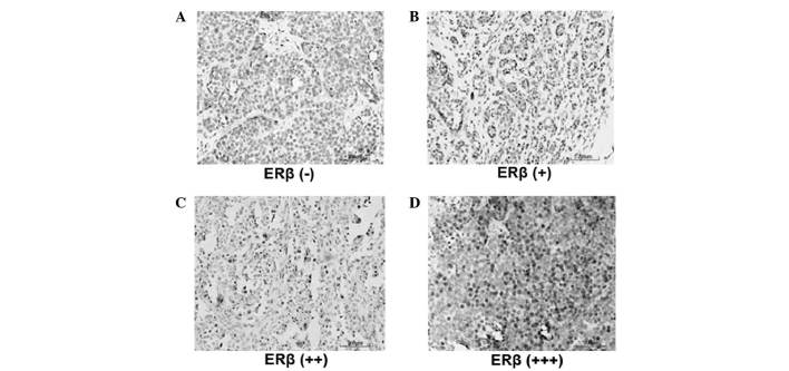
Figure 1.
Expression levels of ERβ in postmenopausal ERα-positive breast cancer patients. Immunohistochemistry was performed to detect the expression levels of ERβ. Representative immunohistochemical results are shown. Cells exhibiting brown staining were classified as ERβ-positive cells. ERβ-positive cells were counted and the ERβ-positive rate was calculated. (A) ERβ (−), ERβ-positive rate <1%. (B) ERβ (+), ERβ-positive rate 1–10%. (C) ERβ (++), ERβ positive rate 10–70%. (D) ERβ (+++), ERβ-positive rate >70%. ER, estrogen receptor.

