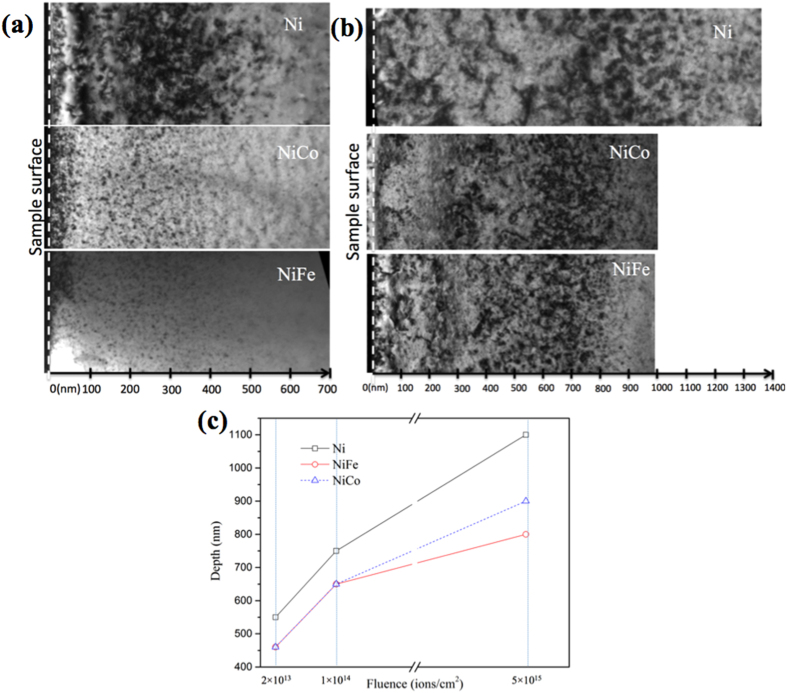
Figure 1. Defect distributions in nickel, NiCo, and NiFe after 3 MeV Au ion irradiation showing the damage range increases with increasing ion fluences and stretches deeper in nickel than in NiCo and NiFe.
Bright-field cross-sectional TEM images (g = [200]) of the samples irradiated to (a) 2 × 10¹³/cm2 and (b) 5 × 10¹⁵/cm2. (c) Plots of the composition and dose dependence of the damage ranges in nickel, NiCo and NiFe irradiated by 3 MeV Au ions to 2 × 10¹³/cm2, 1 × 10¹⁴/cm2 and 5 × 10¹⁵/cm2.

