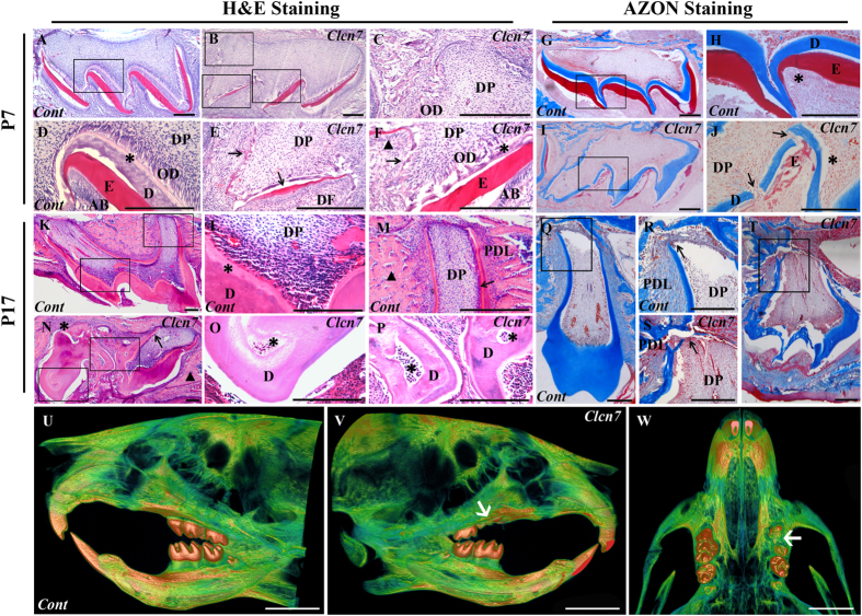
Figure 1. Histological and micro-CT characteristics of tooth germs treated with CS-siRNA particles.
(A–F,K~P) H&E staining; (G~J,Q~T) AZON staining. The molars were harvested and sectioned at day 7 (P7) (A~J) and day 17 (P17) (K~T) from mice treated by control CS-siRNA or CS-Clcn7-siRNA. The P7 molar treated with CS-Clcn7-siRNA showed malformation of enamel and dentin (B,C,E,F). The integrity and continuity of enamel was interrupted (E,F) black arrow: enamel; black triangle: ameloblasts. (I,J). Dentin became disrupted and uneven (J, black arrow) and the boundary between dentin and predentin was indiscernible (F, asterisk), and the disorganized odontoblasts were found (J, asterisk). In P17 CS-Clcn7-siRNA group, the tooth was not fully erupted (N,T). The malformed tooth showed the abnormal cusps and short roots (N, black arrow; T) or disappeared root (N, asterisk). Abnormal and irregular fibrous tissues surrounded tooth instead of bone tissue (N, black triangle). The disorganized dentin and pulp cavity were observed (O,P asterisk). The shape of the tooth crown was irregular and the root dentin was disrupted (S, black arrow) and uneven. The continuity and integrity of cervical loop and Hertwig’s epithelial root sheath was disappeared (S, black arrow). Figure (D,C,E,F,H,J,L,M,O,P,R,S) are partial enlargement of figure (A,B,G,I,K,N,Q,T), respectively. Cont = CS-negative-siRNA treated tooth; Clcn7 = CS-Clcn7-siRNA treated tooth. OD = odontoblasts; DP = dental pulp; DF = dental follicle; E = enamel; AB = ameloblasts; D = dentin; PDL = periodontal ligament. Scale bars = 200 μm. (U~W) Micro-CT scanning image of mouse head. U. Lingual view of the control side treated with CS-negative-siRNA nanoparticle. (V) Lingual view of the experimental side treated with CS-Clcn7-siRNA nanoparticle. W. Palatal view of the maxilla. The left first molar was impacted and lost its regular crown shape and cusps (white arrow). Cont = CS-negative-siRNA treated group; Clcn7 = CS-Clcn7-siRNA treated group. Scale bars = 2 mm.

