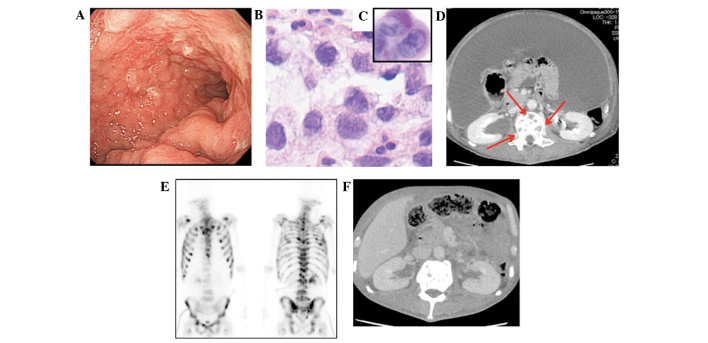
Figure 1.
Clinicopathological features of case 1. (A) Endoscopic view. (B) A pathological view of a gastric tumor. (C) Cytology of ascites. (D) CT on day 2 following the initial treatment. Red arrow indicates bone metastases (osteolytic region). (E) Bone scintigram. (F) CT on day 68. CT, computed tomography.

