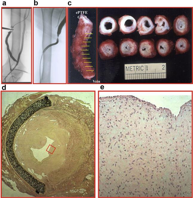
Figure 1.
(a, b) Fistulograms showing a stenosis at the polytetrafluoroethylene graft anastomosis to the basilic vein. (c) A gross specimen from a different patient showing thickening of the vein to graft anastomosis. Hematoxylin and eosin stain (d) at 10× magnification demonstrating a thickened neointima and (e) at 40× magnification (red box in d) showing increased cellular proliferation. ePTFE, expanded polytetrafluoro-ethylene.

