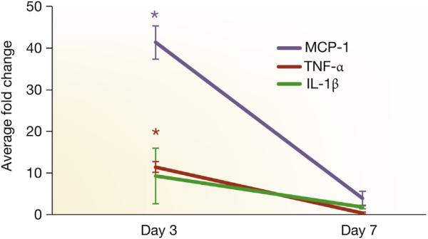
Figure 3. TNF-α, MCP-1, and IL-1β expression by qRT-PCR.
Tissue necrosis factor-alpha (TNF-α), monocyte chemoattractant protein-1 (MCP-1), and interlukin-1 beta (IL-1β) expression by quantitative reverse transcriptase polymerase chain reaction (qRT-PCR) in graft veins and control veins at 3 and 7 days after arteriovenous fistula placement in mice with established chronic kidney disease. There is a significant increase in the mean TNF-α, MCP-1, and IL-1β expression at 3 days in graft veins when compared with control veins (P < 0.05). Each bar shows the mean ± SEM of 3 samples per group. Two-way analysis of variance with Student t test with post hoc Bonferroni correction was performed. *P < 0.05.

