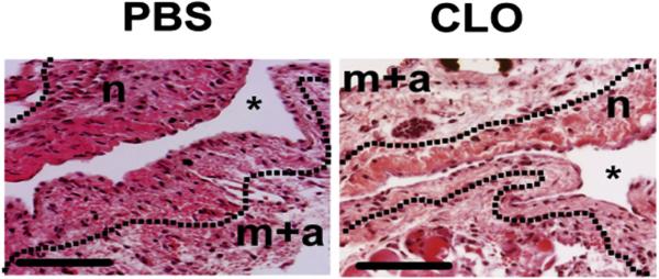
Figure 6. H&E staining of outflow veins removed from animals treated with PBS and CLO at day 14.
Hematoxylin and eosin (H&E) staining of outflow veins removed from PBS-treated and CLO-treated animals at day 14 after arteriovenous fistula placement. There is a reduction in the neointima (n) of the CLO-treated animals when compared with PBS control animals. Asterisk (*) shows the lumen of the vessel. Scale bars = 50 μM. CLO, clodronate; m + a, media/adventitia; PBS, phosphate-buffered saline.

