Fig. 3.
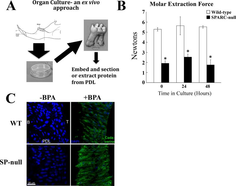
Organ cultures of WT and SPARC-null PDL maintain respective mechanical strength and BPA incorporation by transglutaminase. (A) To further characterize WT and SPARC-null PDL, an organ culture system was developed, as depicted. WT and SPARC-null mandibles were removed and cultured exvivo in growth media as described in Materials and Methods. (B) Mechanical testing of jaws incubated for 24 or 48 hours was carried out to establish that PDL maintained structure and integrity in organ culture. For comparison, the 0 hour time in Fig. 1 is provided. No significant differences in the molar extraction force were detected after 24 or 48 hours of culture within each genotype versus time 0. The molars of SPARC-null teeth continued to require reduced extraction forces versus that of WT teeth. *p < 0.05 between WT and SPARC-null at each isolated time point, by Student's t test. (C). Jaws incubated without BPA (–BPA), a pseudosubstrate of transglutaminase, and with BPA (+BPA), were fixed and sectioned. Staining with avidin-FITC demonstrated BPA incorporation (green) in both WT and SPARC-null PDL with no detectable localization in vehicle control samples (–BPA). SPARC-null sections had increased immunofluorescence intensity, in comparison to sections from WT PDL. DAPI stain of nuclei is shown as blue. Bar = 20 μm. SPARC = secreted protein acidic and rich in cysteine; PDL = periodontal ligament; B = bone; T = tooth; BPA = 5-(Biotinamido) pentylamine.

