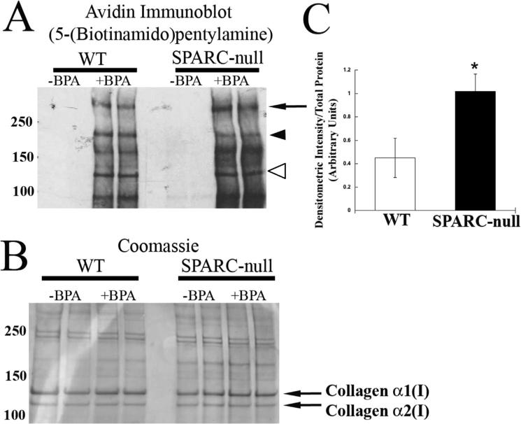
Fig. 4.
Transglutaminase-mediated BPA incorporation is increased in proteins extracted from SPARC-null PDL. (A) Western blot of SDS-PAGE separated PDL protein probed with avidin antibody to detect BPA incorporation. The arrowhead indicates a protein with increased BPA incorporation in a WT PDL, whereas the arrow indicates a protein band that has increased BPA incorporation in SPARC-null extracts. Open arrowhead indicates protein band with migration consistent with that of collagen α1(I). (B) Coomassie-blue–stained gel of proteins loaded in A to demonstrate equal loading of protein. (C) Proteins from SPARC-null PDL have increased incorporation of BPA, as quantified in independent organ culture experiments (n = 4). *p < 0.05 between WT and SPARC-null. SPARC = secreted protein acidic and rich in cysteine; PDL = periodontal ligament; B = bone; T = tooth; BPA = 5-(Biotinamido) pentylamine.

