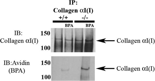
Fig. 5.
Collagen I pulled down from SPARC-null PDL proteins demonstrated increased BPA incorporation versus that in WT PDL extracts. Collagen α1 (I) pulled down from protein extracted from WT and SPARC-null PDL contained BPA-labeled collagen α1(I) (top). SPARC-null PDL collagen α1(I) exhibited increased BPA incorporation in comparison to WT PDL, when equal amounts of starting protein were used. Avidin-HRP Western blots of SPARC-null collagen α1(I) immunoprecipitation also revealed a secondary band, collagen α2(I), labeled with BPA, that was not detected in WT collagen α1(I) pull-downs. The results shown are representative of four independent experiments. BPA = 5-(Biotinamido) pentylamine; IP = immunoprecipitation; IB = immunoblot; HRP = horseradish peroxidase.

