
Figure 2.
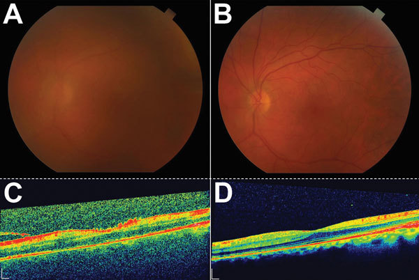
Color fundus and optical coherence tomography (OCT) images during active uveitis and after resolution for a physician from the United States who contracted Ebola virus disease in Liberia and had eye inflammation develop during convalescence. A) Color fundus image of the left eye showing a hazy view to the posterior pole during active uveitis (standardization of uveitis nomenclature classification grade 2–3). B) Color fundus image of the left eye showing a clear view to the posterior pole after resolution of uveitis. C) OCT of macula showing vitreous debris and small particles in a line of vitreous strands, consistent with inflammatory debris. D) OCT of macula showing resolution of vitreous and inflammatory debris. Scale bars indicate 200 μm.